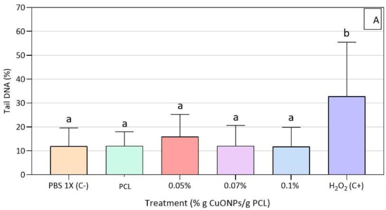
Figure 7.
Genotoxicity results for HFF-1 cells exposed to neat PCL and active PCL films. (A): Genotoxicity assay on HFF-1 cells after 3 h of exposure to the active extracts, (B): HFF-1 cells with PCL + 0.07% CuONPs´ extract, (C): HFF-1 cells with DMEM, (D): HFF-1 cells with H2O2 (80 μM in PBS 1X). Mean values with different letters represent significant differences (p < 0.05) among the samples as determined with a one-way analysis of variance (ANOVA) and Tukey’s multiple comparison tests.


