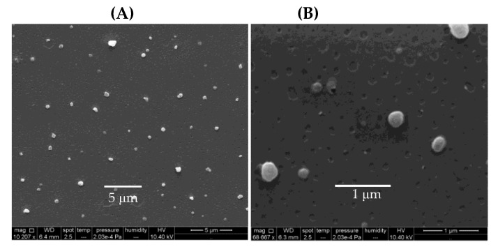
Figure 9.
Scanning electron micrographs of PDDA/OVA NPs assembled at 0.1 mg·mL−1 OVA and 0.01 mg·mL−1 PDDA obtained under low (A) and high magnification (B). At the low PDDA dose employed, PDDA cytotoxicity was not significant against cells in culture. Reprinted from [65].

