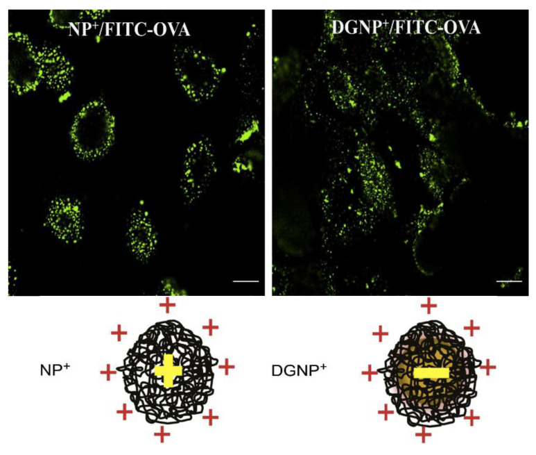
Figure 15.
Comparison of ovalbumin (OVA) intracellular delivery by maltodextrin porous nanoparticles with outer and inner cationic charges (NP+) or outer cationic/inner anionic charges (DGNP+) using confocal microscopy to visualize OVA labeled with fluorescent markers (FITC-OVA). NP+ and DGNP+ were loaded with FITC–OVA and incubated for different periods with 16HBE cells. After 30 min incubation, cells were washed with PBS and fixed with 4% PAF. Intracellular FITC–OVA was visualized by confocal microscopy. Scale bar = 10 μm. Adapted from [244] with permission from Elsevier, Copyright 2012.

