
Figure 6. DNA methylation analysis of stem cells at different developmental stages.
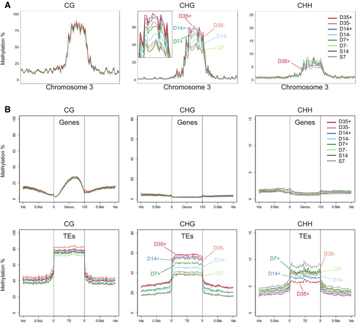
- CG, CHG, and CHH methylation at chromosome 3 in stem and non‐stem cells. The inset for CHG methylation shows a magnification of the pericentromeric region.
- Metaplots of DNA methylation at CG, CHG, and CHH sites for genes (top) and TEs (bottom). + = stem cells; − = non‐stem cells, D7/14/35 = nuclei from 7/14/35‐day-old plants, S7/14 = nuclei from 7/14-day‐old above‐ground seedlings.