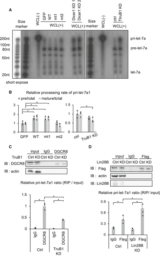
In vitro processing assay for RI‐labeled pri-let‐7a1. Autoradiographs of gels showing pri‐let-7a1 treated with whole cell lysate (WCL) from HEK‐293FT cells transfected with GFP, TruB1, mt1, or mt2 (overexpression, left), and TruB1 KD or ctrl (siRNA, right). Dicer KD was used to show the height of pre‐let-7a1.
RI intensities of (A) were quantified and normalized to ctrl. Relative processing rate of pri‐let-7a1 into pre‐let-7a1 and mature-let‐7a1 are shown. Significance was assessed using 2‐tailed Student's t‐test, < 0.05*.
RIP assay of pri‐let-7a1 and DGCR8 from HEK‐293FT cells. Western blotting for input or immunoprecipitate (IP) using anti‐DGCR8 antibody and anti‐actin antibody are shown on top. RNA was extracted from IP material and analyzed by qRT–PCR (bottom). Significance was assessed using 2‐tailed Student's t‐test, < 0.05*.
RIP assay of pri‐let-7a1 and Flag‐tagged TruB1 from HEK‐293FT cells with Lin28B KD or ctrl (siRNA). Western blotting for input or IP material using anti‐Flag antibody, anti‐Lin28B antibody, and anti‐actin antibody are shown on top. RNA was extracted from IP material and analyzed by qRT–PCR (bottom). Significance was assessed using 2‐tailed Student's t‐test, < 0.05*.
Data information: All experiments were performed in triplicate. Error bars show SD.

