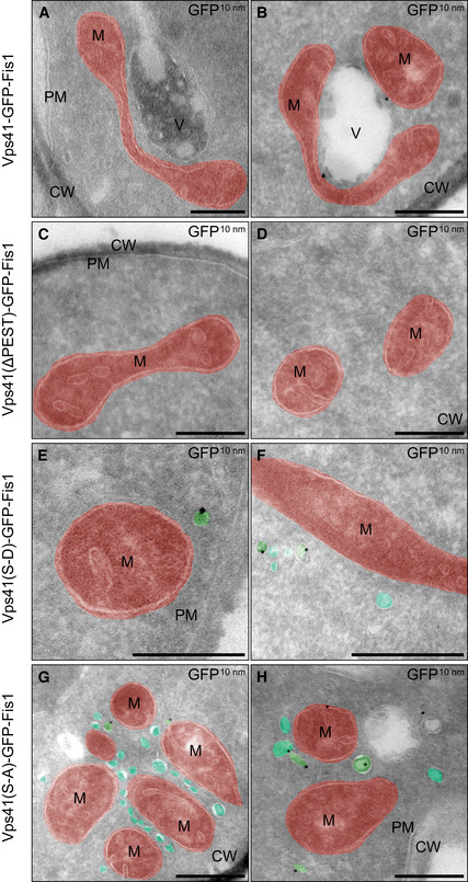
Figure 3. Ultrastructural analysis and immunolabeling of cells expressing mitochondrially anchored Vps41.

-
A–HIndicated strains were grown in YPG for expression of Vps41‐GFP-Fis constructs. Two representative images are shown for each strain. Mitochondria (M) are highlighted in red and vesicular structures in teal (unlabeled for VPS41‐GFP-Fis) or green (unlabeled for VPS41‐GFP-Fis). Cell wall (CW), plasma membrane (PM), and vacuoles (V) are indicated. Scale bars, 250 nm.
