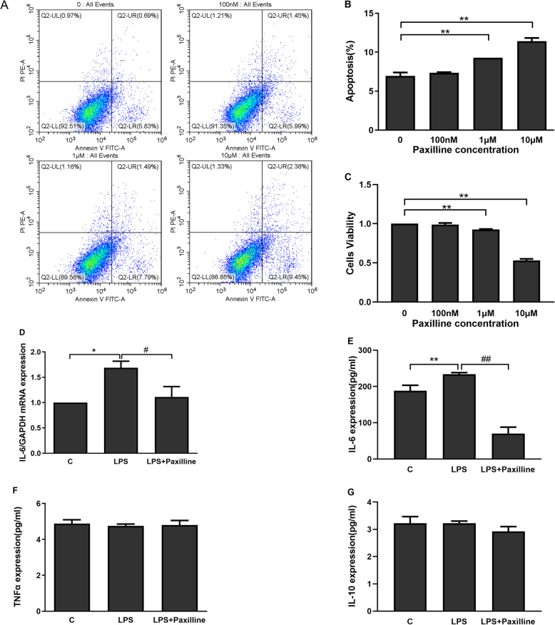
Fig. 4.
BKCa channels in WJ-MSCs induce inflammatory cytokine expression. a, b The percentage of apoptotic WJ-MSCs was analysed using flow cytometry. c The viability of WJ-MSCs was determined by measuring the absorbance at 450 nm using a microplate reader. WJ-MSCs were stimulated with LPS (50 ng/ml) alone or in combination with paxilline (100 nM) for 24 h. d, e The expression of the IL-6 mRNA was analysed using real-time PCR (d), and the IL-6 protein concentration was measured using an ELISA (e). f, g The TNF-α (f) and IL-10 (g) concentrations were measured using ELISAs. The data are presented as the mean ± S.E.M. from three independent experiments. *p < 0.05 and **p < 0.01 compared with the C group or 0 group; #p < 0.05 and ##p < 0.01 compared with the LPS group

