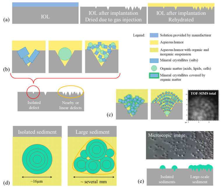
Figure 9.
Diagrams illustrating the probable mechanism of sediment formation on IOL surface. (a) Scheme of lens cross sections at different stages presented in the following order: stored in the liquid foreseen by the manufacturer, after contact with the air bubble after DSAEK procedure, and after rehydration in eye. The probable origins of stages of sediment growth are shown (b) based on the literature and (c) proposed in this work. The sediment expansion was illustrated on scheme (d) top view and (e) cross-section view, combined with microscopic image and TOF–SIMS distribution maps of all detected ions.

