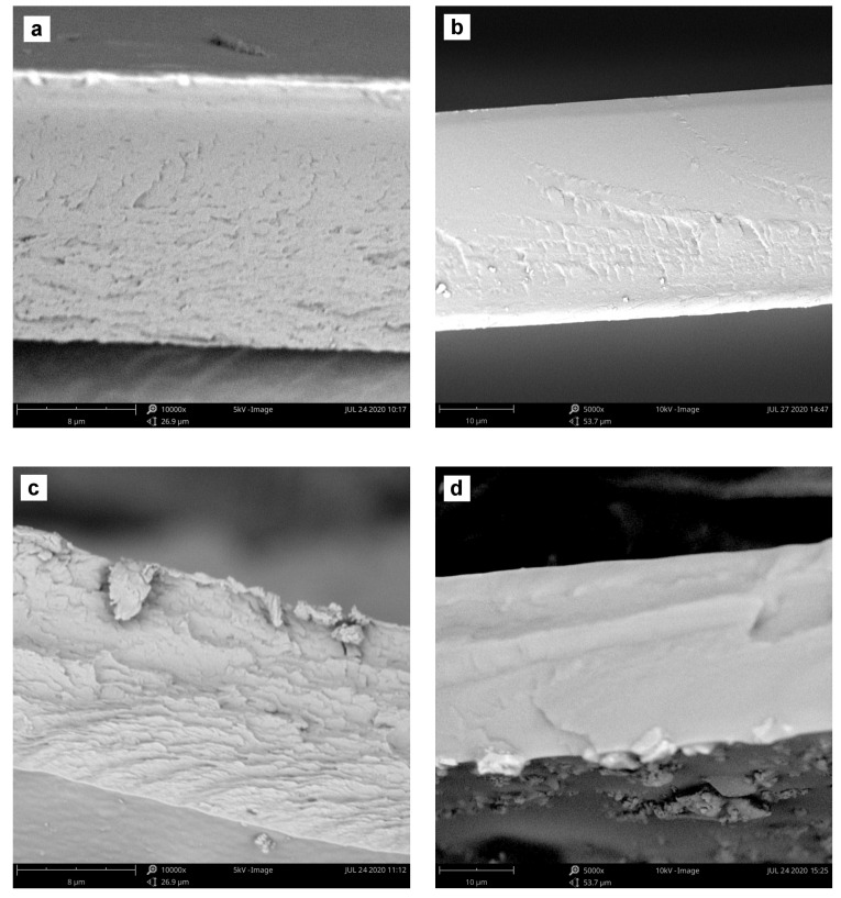
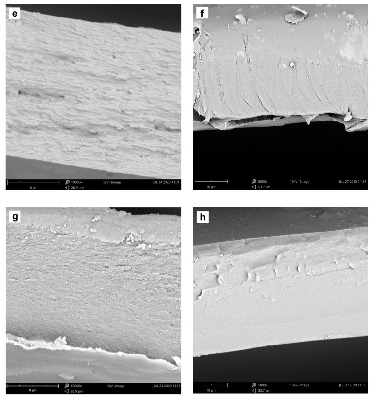
Figure 5.
SEM images of the cross-sectional view of ALG (left column) and PVA (right column) membranes filled with 15 wt% of hematite, magnetite or iron(III) acetylacetonate, respectively. Samples were coated with 5 nm layer of Au. Magnification and acceleration voltage: 10,000× and 5 kV (ALG), and 5000× and 10 kV (PVA). (a) plain ALG; (b) plain PVA; (c) ALG_Fe2O3; (d) PVA_Fe2O3; (e) ALG_Fe3O4; (f) PVA_Fe3O4; (g) ALG_Fe(acac)3 and (h) PVA_Fe(acac)3.


