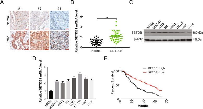
Fig. 1.
SETDB1 was upregulated in glioblastoma tissues and corelative with poor tumor progression. a Representative IHC staining of SETDB1 protein expression in glioblastoma tumor tissues (T) and adjacent normal tissue (N) of three patients. Scale bar: 50 μm. b mRNA level of SETDB1 was analyzed by real-time PCR in glioblastoma tumor tissues (T) and adjacent normal tissue (N) of patients. c Western blotting of SETDB1 expression in NHAs and indicated glioblastoma cells. d mRNA level of SETDB1 was analyzed by real-time PCR in NHAs and indicated glioblastoma cells. e Kaplan-Meier survival analysis indicated that glioblastoma patients with high expression of SETDB1 had worse relapse-free survival

