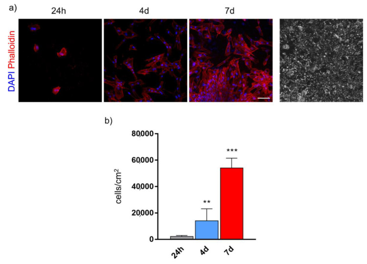
Figure 3.
Cell morphology (a) and proliferation (b) of human dental pulp stem cells (hDPSCs) cultured on BGMS10 surfaces at different time points. Immunofluorescence analysis shows representative images of hDPSCs at 24 h, 4 days and 7 days. On the right, BGMS10 surface is shown. ** p < 0.01 vs. 24 h, *** p < 0.001 7 days vs. 4 days and vs. 24 h. Scale bar: 50 μm.

