FIGURE 1.

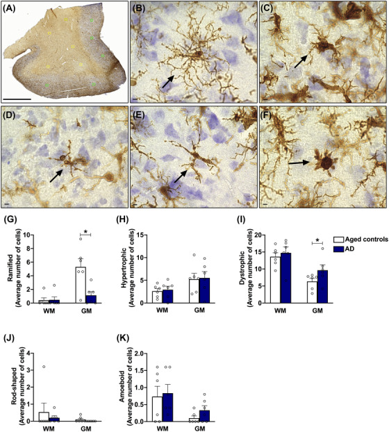
Microglia morphologies evaluated in the posterior cingulate cortex, and microglia cell counts in the Alzheimer's disease (AD) brain. A, Characterization of microglia was performed in five random 250 × 250 micron boxes in the white matter and five random 250 × 250 micron boxes in the gray matter for each tissue section Scale: 3 μm. Five microglia morphologies were analyzed: (B) ramified microglia, which have thin, branched processes to actively “survey” changes in their environment; (C) hypertrophic microglia (also known as activated microglia), which may have enlarged, short processes and thicker bodies; (D) dystrophic microglia, that present fragmented or “beaded” processes possibly due to microglial dysfunction as a result of aging; (E) rod‐shaped microglia, that have elongated nuclei, few processes, and are most notable in chronic disorders; (F) amoeboid microglia, which have a round body, lack processes, and appear in response to acute destruction of central nervous system tissue. Scale: 5 μm. G–K, In the white matter, no significant changes were observed in any of the microglial phenotypes analyzed. In the gray matter, a significant reduction was observed in the number of ramified cells in the AD group, while the number of dystrophic cells was increased, compared to aged controls. Data are presented as mean ± standard error of the mean, * P ≤ 0.05 (Student's t test)
