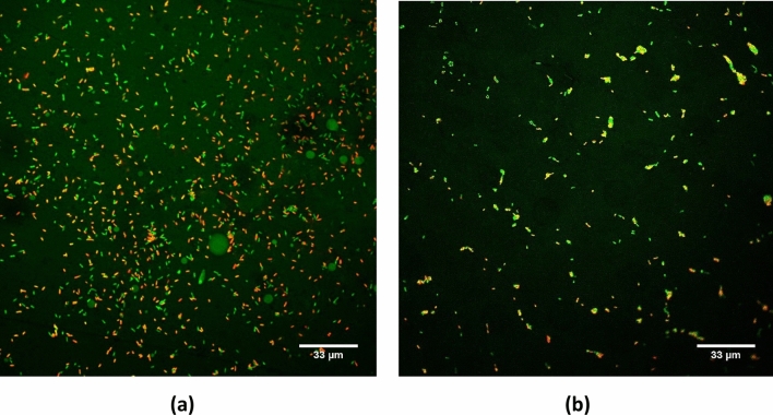
Figure 3.
Confocal laser scanning microscope images of E. coli biofilm on glass slide (a) non-treated and following (b) 15 min treatment of airborne ultrasound. The cells were stained with STYO 9 (green fluorescence, live cells)/PI (red/yellow, dead cells). All images were compiled, and noise reduction filter was applied in the Olympus Fluoview FV1000 software (version 4.1.1.5. https://www.olympus-lifescience.com/en/support/downloads/ ).

