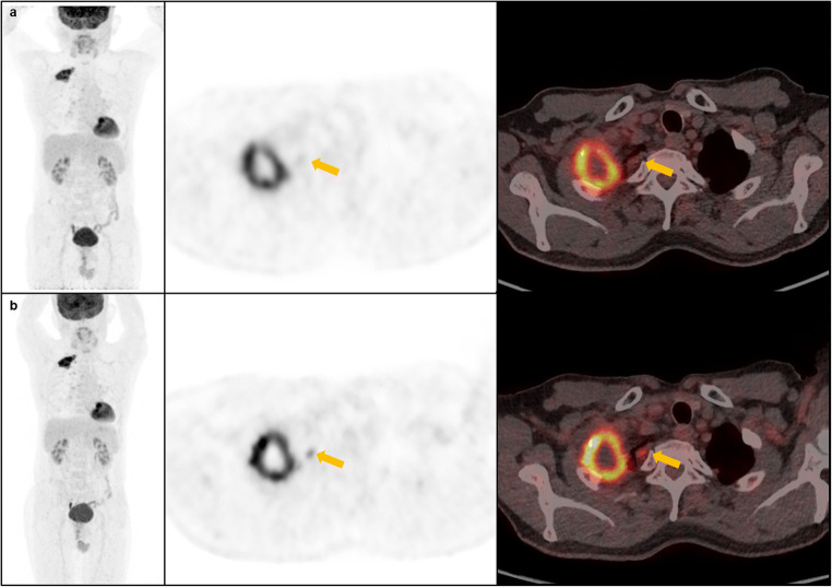
Fig. 4.
Representative MIP, axial PET and fusion PET/CT images (from left to right) acquired on the standard PET/CT (a, upper row) and digital PET/CT (b, lower row) of a 68-year-old male patient with lung cancer. A small pleural lesion (yellow arrows) found on digital PET/CT that did not appear as such on standard PET/CT images

