Fig. 1.
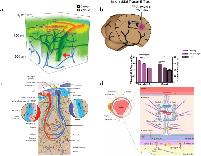
Interstitial fluid (ISF)-cerebrospinal fluid (CSF) space connection. Glymphatic hypothesis and their experimental evidence in rodents. a Via cisterna magna cannula, during sleep at noon FITC-dextran (3 kDa, green), was given, and after being awakened 30 min later, Texas-red-dextran (red) was given. On in vivo two-photon microscopy, deeper/broader fluorescence during sleep and reduced fluorescence after being awakened were interpreted to represent the CSF influx of intra-cisternal administered small molecules. Reprinted with permission from Xie et al. [18]. b To ISF space of caudate nucleus via guide cannula, I-125 amyloid-beta and C-14 inulin were injected, and 60 min later, the radioactivity of the brain was measured to yield (100%-brain recovery) as percent clearance. ISF clearance decreased upon aging. Reprinted with permission from Kress et al. [19]. c ISF-CSF space is communicating through the basement membrane of the endfeet of astrocytes surrounding brain blood vessels through perivascular spaces (periarteriolar and perivenular spaces are well-documented and pericapillary space is supposed between basement membranes of astrocytic endfeet and vascular endothelial cells. Pericytes are located in this space. Reprinted with permission from Da Mesquita et al. [17]. d Schematic magnification of endothelial and astrocytic basement membranes. Reprinted with permission from Mastorakos and McGavern [20]. If periarteriolar and perivenular spaces filled with CSF are supposed to be connected with pericapillary spaces between basement membranes, the CSF network around parenchymal vessels was expressed as a circuit of paravascular spaces running CSF flow throughout brain parenchyma. Reprinted with permission from Coles et al. [21]

