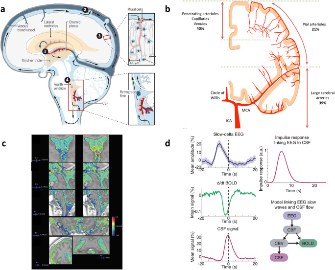
Fig. 4.
CSF circulation in relation to parenchymal vessels. CSF was once considered to circulate but in fact, not. a Classic understanding of CSF flow produced from ventricles (lateral, third, and fourth), flowing out to cisterns and going down to the spinal canal and over the cord and returning to basal and inter- or over the hemispheric subarachnoid spaces to reach arachnoid villi to be absorbed. In this picture, the flow between the third and fourth ventricle is said to be found bidirectional. Aqueduct connecting third and fourth ventricles are open with this bidirectional flow. Reprinted with permission from Grubb and Lauritzen [42]. b Internal carotid artery supplies wider than anterior two-thirds of both cerebral hemispheres. Large or small, vessels outside of parenchyma are responsible for 60% of total resistance (39% + 21%) and penetrating arterioles and venules for the remaining 40%. Reprinted with permission from Iadecola [43]. c On 3D phase-contrast MRI images, CSF flow was found not to be circulatory, but complex, accelerating/decelerating, unstable, and augmented in the center of the cranial cavity of subarachnoid space, and third/fourth ventricles [44, 45]. Reprinted with permission from Matsumae et al. [44]. d On simultaneous measurements of EEG, BOLD on fMRI, and CSF flow dynamics imaging, during NREM sleep, slow delta wave (increased amplitude) preceded BOLD changes (decreased signal) which also preceded CSF flow oscillatory changes. NREM sleep with its impact upon hemodynamic status results macroscopic oscillations in fourth ventricle CSF. Reprinted with permission from Fultz et al. [46]

