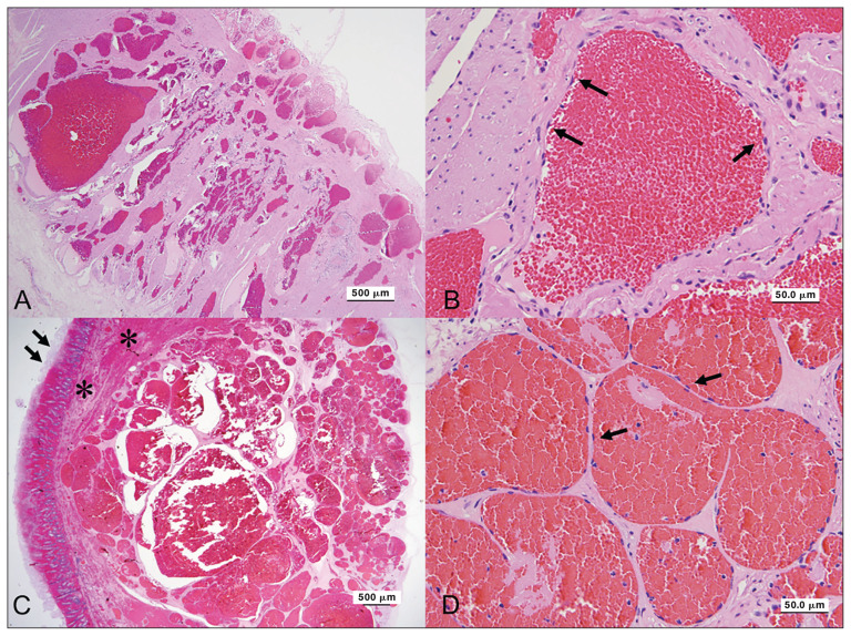
Figure 1.
Angiomatosis in the jejunum of a horse. A — The lesion in the grossly normal jejunum consists of numerous variable-sized blood vessels mainly in the muscular layer. H&E stain. Scale bar = 500 μm. B — In the grossly normal jejunum, the vascular channels are lined by well-differentiated flattened endothelial cells (arrows). H&E stain. Scale bar = 50 μm. C — The lesion in the grossly normal jejunum consists of numerous variable-sized blood vessels mainly in the muscular layer (arrows). The mucosal layers contain abundant congestion and hemorrhage with mucosal necrosis (asterisks). H&E stain. Scale bar = 500 μm. D — In the grossly abnormal jejunum, the vascular channels are lined by well-differentiated flattened endothelial cells (arrows). H&E stain. Scale bar = 50 μm.

