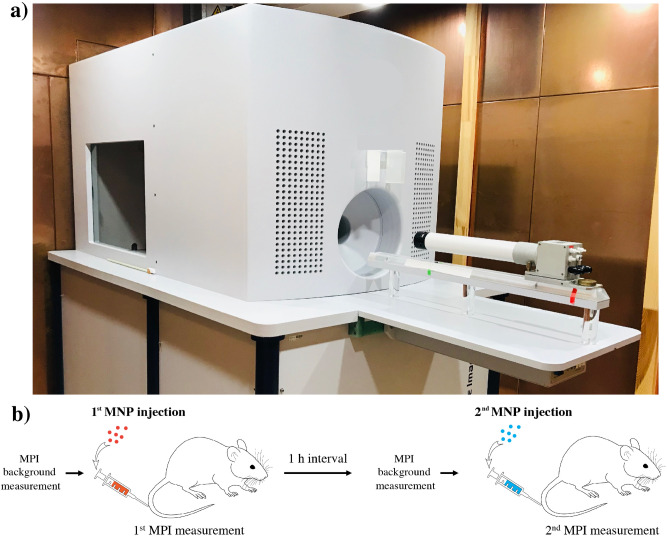
Figure 6.
Preclinical MPI scanner (a) and diagram of experimental workflow (b). (a) Preclinical MPI scanner (MPI 25/20 FF, Bruker BioSpin) in a copper-shielded cabinet to suppress interferences from external electromagnetic fields. (b) Diagram of experimental workflow. First, background MPI signal is acquired, followed by IV injection of one of the MNP and MPI scanning for several minutes. After 1 h (no signals from MNP detectable in blood vessels), the second tracer, was injected intravenously. The order of MNP type administration was random.

