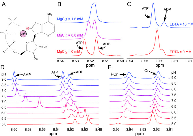
Figure 1.
Defining the conditions to allow the quantification of purine nucleotides from extracts of cardiac muscle. (A) Schematic of ATP with Mg2+ ion. The proton used for distinct quantification is highlighted in red. (B) The effect of Mg2+ ion concentration in equimolar concentrations of ATP and ADP. The presence of Mg2+ alters the chemical shift of the ATP proton, but not the ADP; resulting in a convergence of these peaks with physiological concentrations of Mg2+. (C) 1H-NMR spectra from polar metabolites extracted from perfused mouse hearts without (red) and with EDTA (blue). In the presence of EDTA, the ATP and ADP peaks are clearly distinct and easily quantifiable. (D) The effect of extract pH on the separation of the purine proton peaks, note the high mobility of the AMP proton. (E) The pH sensitivity creatine (Cr) and phosphocreatine (PCr) protons.

