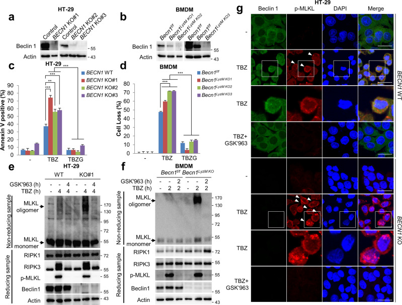
Fig. 5. Beclin 1 knockout promotes necroptosis.
a Beclin 1 protein levels were determined by immunoblotting in Beclin 1 wild type (WT) or knockout (KO) HT-29 cells. b Beclin 1 protein was detected by immunoblotting in Becn1F/F and Becn1LyzMKO BMDM. c, e Beclin 1 WT or KO HT-29 cells were treated with TBZ in the presence or absence of GSK'963 (TBZG) for 4 h. After treatment, the cells were stained with annexin V-FITC and 7-AAD for 20 min, and then analysed via flow cytometry (c). To analyse MLKL oligomerisation, the cells were lysed under non-reducing conditions for MLKL oligomerisation or under reducing conditions, and then analysed by immunoblotting (e). Flow cytometry data are the means ± S.D., n = 3, with **P < 0.01 and ***P < 0.001 at each point compared to indicated graph with the two-sided Student’s t test (c). d, f Becn1F/F and Becn1LyzMKO BMDMs were treated with 20 ng/mL TNFα, 1 μM BV6, and 20 μM Z-VAD-FMK (TBZ) in the presence or absence of 100 nM GSK’963 (TBZG) for 2 h. After treatment, the cell loss was determined via cell titer-glo (d). The cell loss data are the means ± S.D., n = 3, with ***P < 0.001 at each point compared to indicated graph with the two-sided Student’s t test (d). To analyse MLKL oligomerisation, the cells were lysed under non-reducing conditions for MLKL oligomerisation or under reducing conditions, and then analysed by immunoblotting (f). g Beclin 1 WT and KO HT-29 cells were treated with TBZ in the absence or presence of GSK’963 for 5 h. After treatment, the cells were fixed and stained with the anti-Beclin 1, p-MLKL antibodies, and DAPI. The boxed areas are shown at higher magnification to the below. Scale bars = 20 μm.

