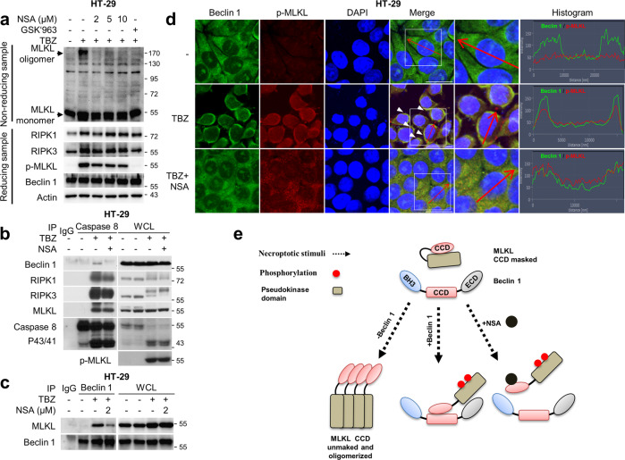
Fig. 7. NSA and Beclin 1 compete for CCD of MLKL.
a HT-29 cells were treated with TBZ in the presence or absence of GSK’963 or NSA. After treatment, cell were lysed under non-reducing conditions for MLKL oligomerisation or under reducing conditions, and then analysed by immunoblotting. b HT-29 cells treated with TBZ in the presence or absence of 5 μM NSA for 5 h. After treatment, the cell lysates were immunoprecipitated using the anti-caspase-8 antibody, and then analysed by immunoblotting. c HT-29 cells treated with TBZ in the presence or absence of NSA were lysed and immunoprecipitated using the anti-Beclin 1 antibody, followed by immunoblotting analysis. d HT-29 cells treated with TBZ in the presence or absence of NSA for 5 h were fixed and stained with the anti-Beclin 1, p-MLKL antibodies, and DAPI. The histogram represents the subcellular integrity of Beclin 1 and p-MLKL of the areas indicated by the red arrows. The boxed areas are shown at higher magnification to the right. Scale bars = 20 μm. e Scheme for the hypothetical model of interaction between Beclin 1 and MLKL with necroptotic stimuli in the presence or absence of NSA.

