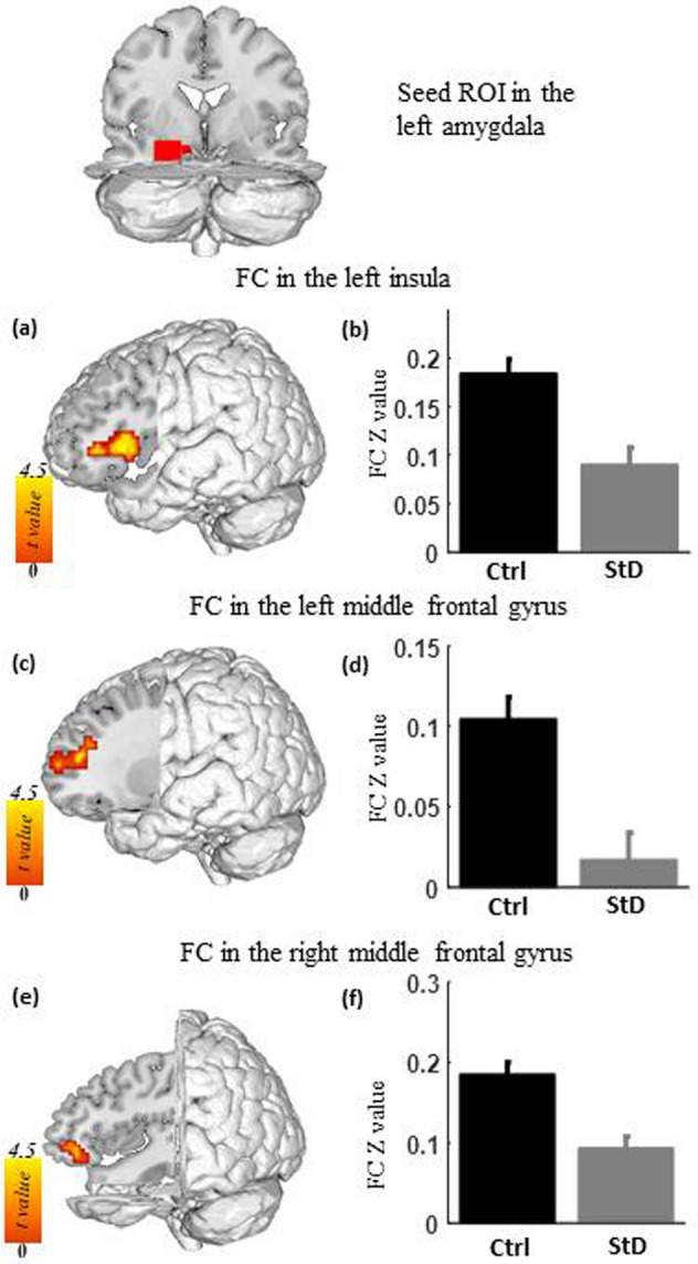
Figure 2.

Functional connectivity of the left amygdala in individuals with StD compared with healthy controls (Ctrl). (a), (c), and (e) Compared with the control, the StD group showed decreased functional connectivity between the left amygdala seed region and the left insula (a), the left middle frontal gyrus (c), and the right middle frontal gyrus (e). (b), (d), and (f) Average functional connectivity of the left insula (b), the left middle frontal gyrus (d), and the right middle frontal gyrus (f) in both groups.
