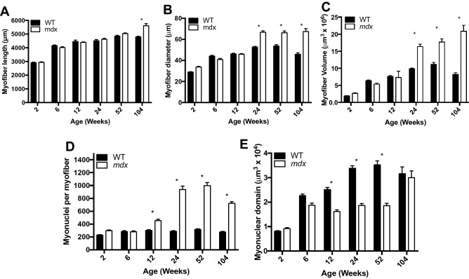
Figure 2.
Mdx EDL myofibers are hypertrophic, hypernucleated and have a higher density of myonuclei relative to WT. (A) The length of mdx myofibers is not different from WT mice until 2 years of age, at which time extensive branching architecture occurs in mdx muscles. (B) The diameter of mdx myofibers is larger than age matched WT starting at 24 weeks and continuing through the lifespan. (C) Mdx myofibers become progressively hypertrophic throughout the lifespan and are not susceptible to atrophy in old age. (D) Mdx myofibers become hypernucleated by 12 weeks of age demonstrating recruitment of new myonuclei prior to the onset of hypertrophy. Hypernucleation increases through the first year of life and remains elevated through the lifespan. (E) The myonuclear domain of mdx mice is smaller than WT from 12 to 52 weeks of age indicating a higher density of myonuclei. N = 3 mice and 30 myofibers per group. Analysis via two-way ANOVA with Tukey post hoc test. * = p < 0.05. All data sets are displayed as mean ± s.e.m.

