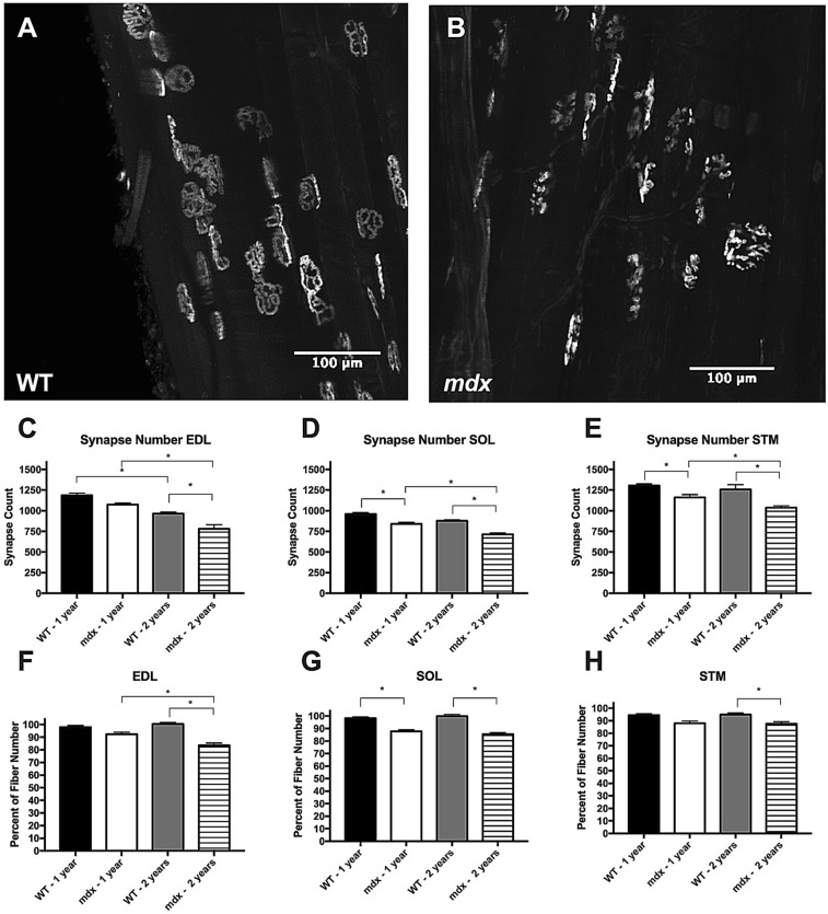
Figure 8.
Mdx mice are susceptible to myofiber loss. (A) BTX labeled endplates from a cleared adult WT EDL muscle. Most endplates display the normal continuous morphology. (B) BTX labeled endplates from a cleared adult mdx EDL muscle. Most endplates display the fragmented morphology. (C) Synapse loss occurs as a function of age in WT and mdx EDL muscles. At 2 years of age, mdx EDL muscles have significantly fewer synapses than age matched WT. (D) Synapse loss occurs as a function of age in mdx SOL muscles and trends towards the same in WT. At 1 and 2 years of age mdx SOL muscles have significantly fewer synapses that age matched WT. (E) Synapse loss occurs as a function of age in mdx STM muscles. At 1 and 2 years of age mdx STM muscles have significantly fewer synapses than age matched WT. (F) Synapse number normalized against group matched myofiber number in the EDL muscle. The ratio of myofiber count to synapse count is significantly lower in aged mdx muscles compared to adult mdx and aged WT groups. (G) Synapse number normalized against group matched myofiber number in the SOL muscle. The ratio of myofiber count to synapse count is significantly lower in mdx muscles compared to age matched WT. (H) Synapse number normalized against group matched myofiber number in the STM muscle. The ratio of myofiber count to synapse count is significantly lower in aged mdx muscles compared aged WT muscles. Analysis via two-way ANOVA with Tukey post hoc test. * = p < 0.05. All data sets are displayed as mean ± s.e.m.

