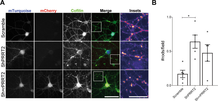
Fig. 5. Knockdown of PRRT2 in hippocampal neurons induces the formation of cofilin-actin rods.
A 7 DIV hippocampal neurons were infected with either mTurquoise-tagged Scramble or ShPRRT2 or co-infected with mTurquoise-ShPRRT2 and rPRRT2-mCherry lentiviral vectors. After 7 days (14 DIV), rods were stained using cofilin antibody and recognized as dense elongated bundles along neurites as shown in the insets. Scale bar: 20 μm (Insets: 25 μm). B Quantitative analysis of the rod density expressed as number of rods per field. Mean ± SEM n. rods/field: Scramble = 0.170 ± 0.05, ShPRRT2 = 0.641 ± 0.09, Sh+rPRRT2 = 0.477 ± 0.12. Data are expressed as mean ± SEM of the number of rods found in each experiment from five independent experiments. For each coverslip at least 20 random fields were acquired. One-way ANOVA/Bonferroni’s tests; *p < 0.05.

