
Figure 2.
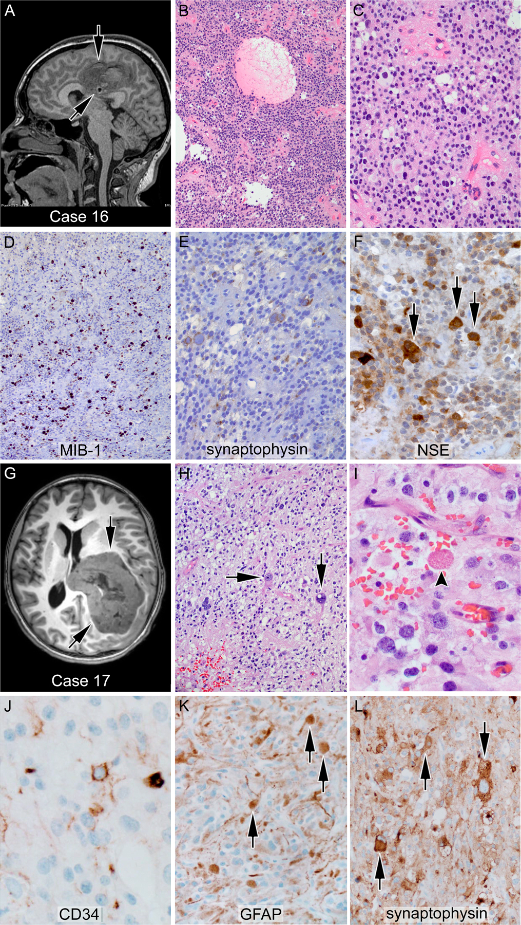
Glioneuronal tumors with CIC-LEUTX alteration; (A-F) case 16—anaplastic ganglioglioma: the tumor showed a population of monotonous cells with round nuclei and a second population of larger ganglionic cells (F, arrows) with positivity for neuronal markers, including synaptophysin and neuron-specific enolase. (G-L) Case 17—anaplastic pleomorphic xanthoastrocytoma: a markedly pleomorphic tumor with large bizarre cells (arrows), occasional xanthomatous cells, and rare eosinophilic granular bodies (I, arrowhead); an arborizing pattern of CD34 positivity was seen (J). The large cells (K, L arrows) are positive for both glial and neuronal markers such as GFAP and synaptophysin, respectively. More than 5/10 high power fields mitoses were found, sufficient for an anaplastic (grade III) diagnosis. A and G represent T2- and T1-weighted magnetic resonance images, respectively, of the corresponding patients’ brains. Arrows indicate the location of the tumor.