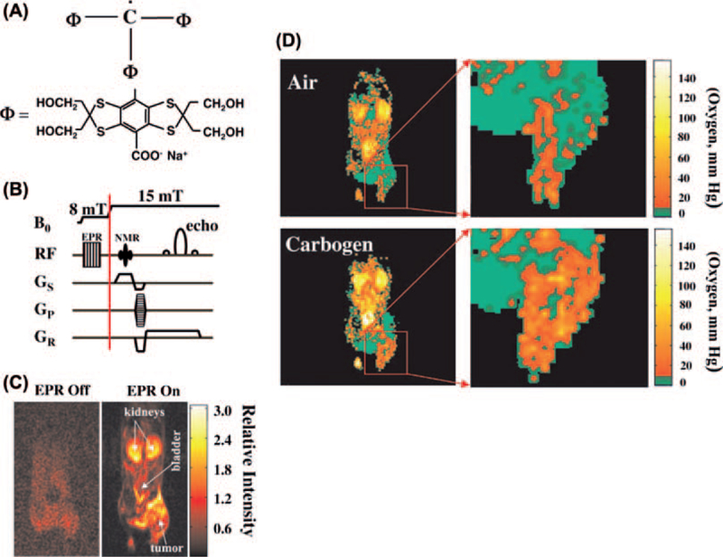
Figure 1.
(A) Structural formula of the electron paramagnetic agent OX063, a trityl radical. (B) Overhauser MRI pulse-sequence diagram showing B0 filed cycling and RF and field-gradient waveforms. (C) Interleaved (“EPR off” and “EPR on”) OMRI images (coronal) of a female C3H mouse, bearing SCC tumor on the right hind leg, demonstrating the Overhauser enhancement. Both images were acquired in the presence of the contrast agent. (D) pO2 images of a mouse with SCC tumor during air breathing (upper) and carbogen breathing (lower). The expanded tumor region is given at the right (see [18]).

