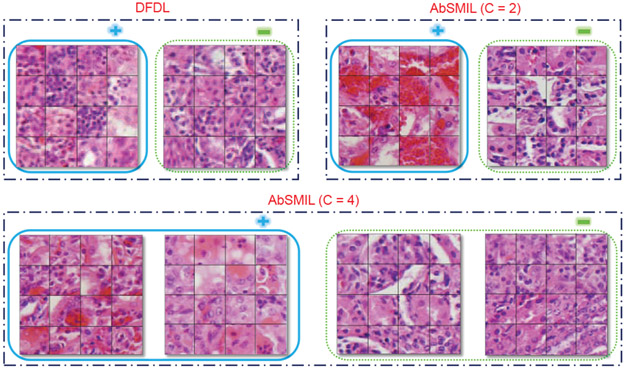
Fig. 6.
Groups of instances in the same class predicted by AbSMIL and DFDL. Cancer groups are in solid blue while normal ones in dotted green. Compared to two DFDL bases, AbSMIL exhibits distinctly different tissue types between the two categories with and can be clustered to distinguish phenomenon in each category with .

