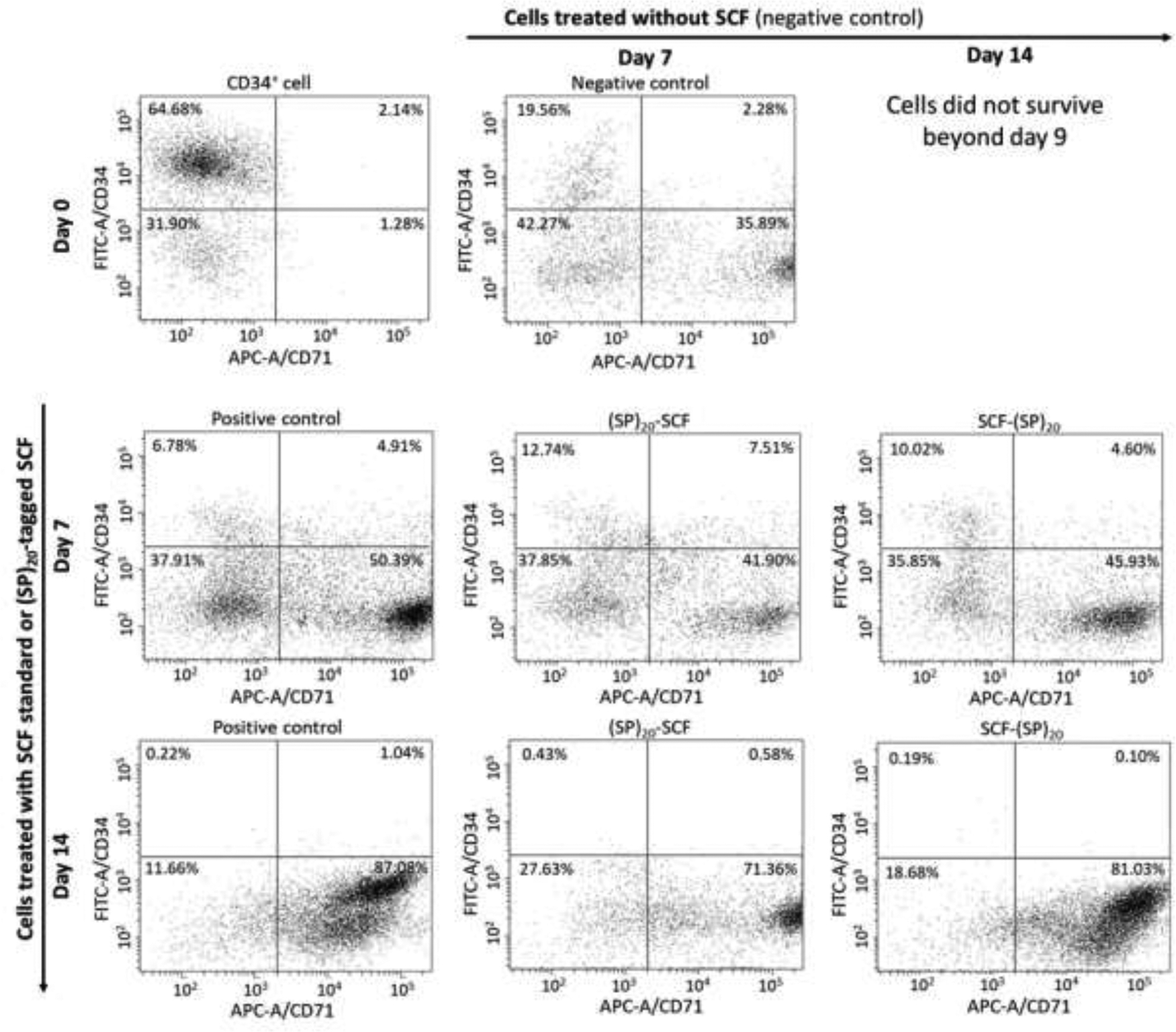
Figure 7. Flow cytometric analysis of the CD34+ cell differentiation after 7 and 14 days of incubation.

The differentiated cells were labelled with a FITC for the CD34 marker and an APC for the CD71 marker. Positive control: CD34+ cells treated with the commercial SCF in combination with the other growth factors; negative control: CD34+ cells treated with the growth factors lacking the SCF; Two sample groups: CD34+ cells treated with the (SP)20-SCF or SCF-(SP)20 in combination with the other growth factors.
