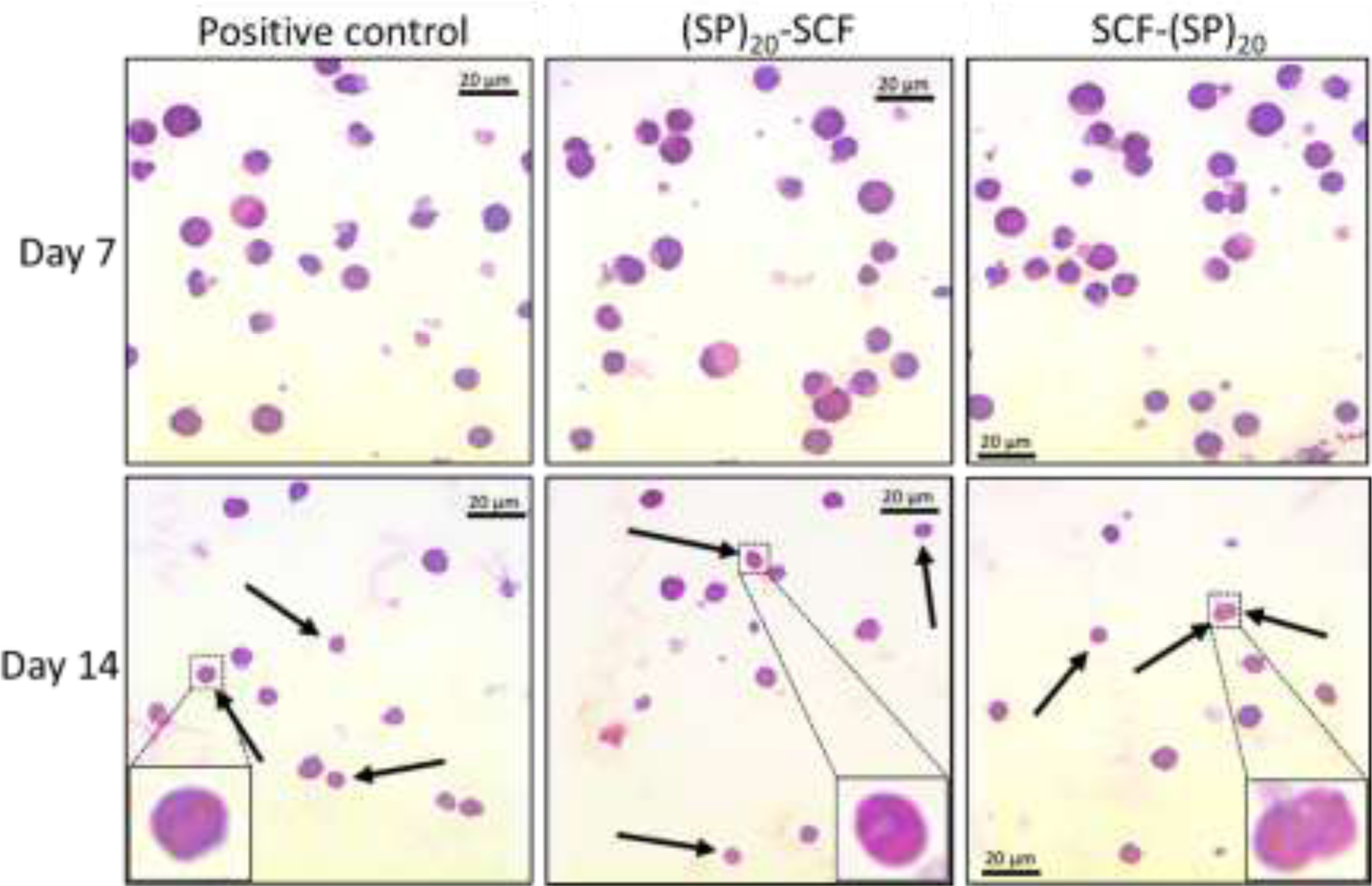
Figure 8. Microscopic images of the differentiating CD34+ cells with a Giemsa staining.

Enucleated RBCs are indicated by the arrows. Some matured RBCs with a donut-like shape are enlarged in the boxes. bar = 20 μm

Enucleated RBCs are indicated by the arrows. Some matured RBCs with a donut-like shape are enlarged in the boxes. bar = 20 μm