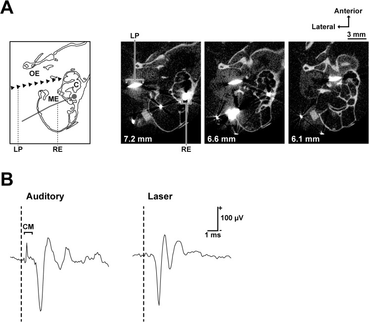
Fig 1. Laser irradiation site and the cochlear responses to laser and auditory stimuli.
(A) Schematic of recording and stimulation configuration (the left-most) and microcomputed tomography images of a stimulated cochlea (horizontal section). The distance from the sagittal suture is stated on the bottom left of each image. The recording electrode (silver wire) and the laser path (tungsten wire) are shown as larger than their actual sizes due to metal artifacts in CT images. (B) Cochlear responses to auditory (100 μs single click with 70 dB peak-to-peak equivalent SPL) and laser (100 μs single pulses, 4.9 mJ/cm2) stimuli. A clear cochlear microphonic (CM) was observed only after the auditory stimulus. The vertical dashed lines show stimulus onset. Abbreviations: OE, outer ear; ME, middle ear; C, cochlea; LP, laser path; RE, recording electrode; CM, cochlear microphonic.

