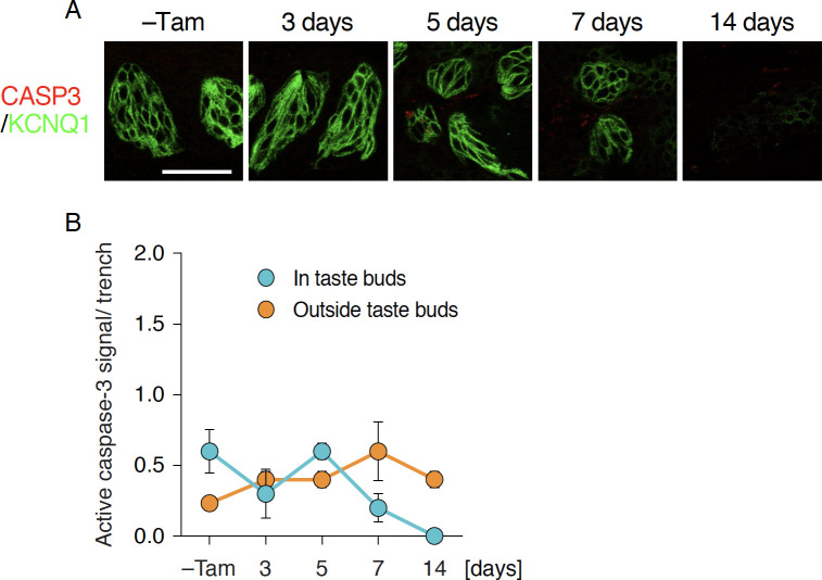
Fig 3. Unaltered apoptotic cell death after Sox2 deletion in epithelial stem/progenitor cells.
A: Double fluorescence immunohistochemical labeling of active CASP3 (red) and KCNQ1 (green) in CvP of Krt5CreERT2/+; Sox2flox/flox mice with and without tamoxifen injection (–Tam, control). Scale bar, 50 μm. B: Quantitative analyses of active CASP3+ cells in (blue) and outside taste buds (orange). Numbers of active CASP3+ cells per trench wall were statistically analyzed using Welch’s ANOVA to evaluate significant change over time (n = 3 at each time point). The data are expressed as the mean ± s.e.m.

