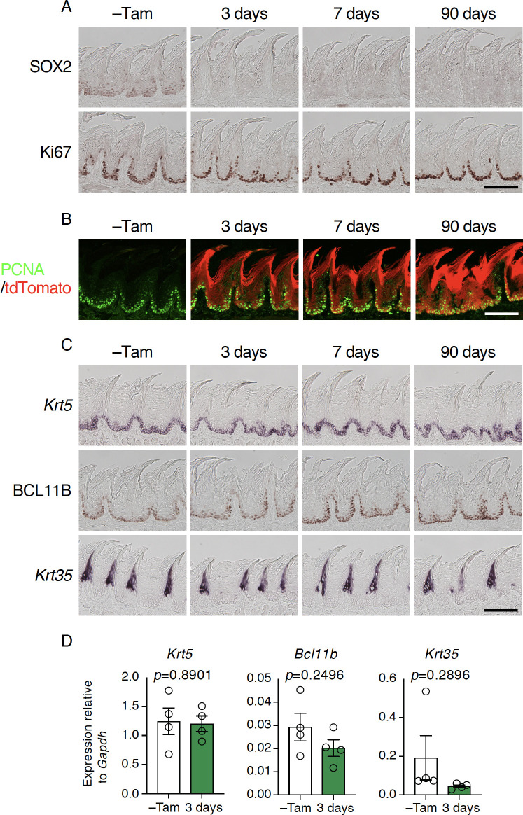
Fig 7. Sox2 is dispensable for the normal turnover of non-gustatory papillary epithelial cells.
A: Immunohistochemical staining of SOX2 (top) and Ki67 (bottom) in filiform papillae (FiP) in the intermolar eminence with and without tamoxifen injection (–Tam, control). B: Lineage tracing by tdTomato induced concurrently with Sox2 deletion in stem/progenitor cells. Immunoreactive signal to PCNA (green) and tdTomato epifluorescence (red) are overlaid. C: Expression of marker genes and protein expressed in epithelial cells at distinct differentiation stages.Mice used are Krt5CreERT2/+; Sox2flox/flox (n = 2 for–Tam, 3 days, and 7 days) and Krt5CreERT2/+; Sox2flox/flox; Rosa26lsl-Tom/+ mice (n = 1 for–Tam, 3 days, and 7 days; n = 3 for 90 days). Scale bars, 50 μm. D: Quantitative PCR analyses to evaluate epithelial cell marker gene expression in FiP in the intermolar eminence. Relative gene expression levels were normalized using Gapdh and statistically evaluated by Welch’s t-test (n = 4 each, Krt5CreERT2/+; Sox2flox/flox mice before and 3 days after tamoxifen injection).

