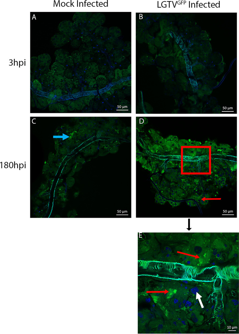
Fig 5. Localization of LGTVGFP in infected SG cultures from male ticks.

Whole mounts of mock- and green fluorescent protein tagged-Langat virus (LGTVGFP)-infected salivary glands (SGs) from male Ixodes scapularis ticks were imaged via confocal microscopy. Each image displays a single SG from a pool of 3 SG pairs per experimental group. A-D images were taken at 40X and insert E was taken at 63X magnification, representing the portion of image D outlined with the red box. Image processing was consistent among respective timepoints. LGTVGFP-infected SGs shows both discrete puncta as well as broad aggregations of GFP expression in granular acini. Red arrows indicate LGTVGFP, blue arrows indicate autofluorescence, and white arrows indicate DAPI stained cell nuclei. Samples from mock-infected organs were shown in order to compare SG autofluorescence.
