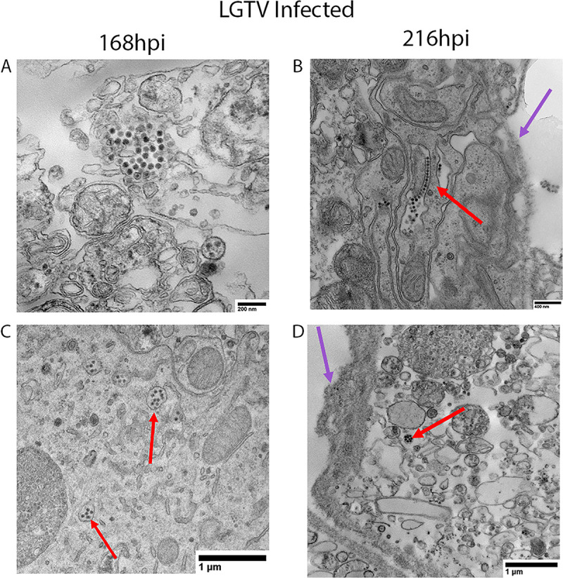
Fig 7. Transmission electron microscopy of LGTV-infected SG cultures from male ticks.

Male Ixodes scapularis salivary gland (SG) culture samples were prepared for transmission electron microscopy at 168 hours post infection (hpi) and 216hpi. Each image displays a single SG from a pool of 3 SG pairs per experimental group. In LGTV-infected samples, virus particles were packaged in single-membrane-bound compartments (A, C, and D) or extracellular (B) and had a diameter of approximately 41-42nm. Red arrows indicate virus particles and purple arrows denote the lumen of the acinus. Panel B displays virus particles that appear to be extracellular.
