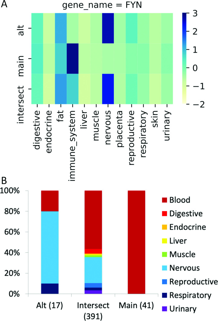
Fig 4. The group-specific splicing event in FYN.
(A) Group-specific distribution of reads that support each side of the FYN splice junction (main, alternative) and those that support the common protein sequence (intersect) coloured by standard deviation from the mean; the darker the colour, the greater the positive standard deviation. The main transcript has more than one standard deviation of reads than the alternative for immune system tissues and the alternative transcript has more than one standard deviation of reads than the main transcript in nervous tissues. (B) The distribution of the PEDs from the proteomics experiments for the alternative and main isoforms (“Alt” and “Main”) and those that map to the remainder of the common amino acid sequence (“Intersect”). The numbers of PEDs that belong to each group are shown in brackets. Fisher tests show that the alternative side of the event is significantly enriched in peptides from nervous tissues, just as in the RNAseq experiment. The main side of the event is significantly enriched in peptides from blood cells. Although both sides of the event are enriched in different tissue groups, FYN does not count twice towards the total of 99 cases in the PGE99 set because the main side of the event is enriched in blood cells.

