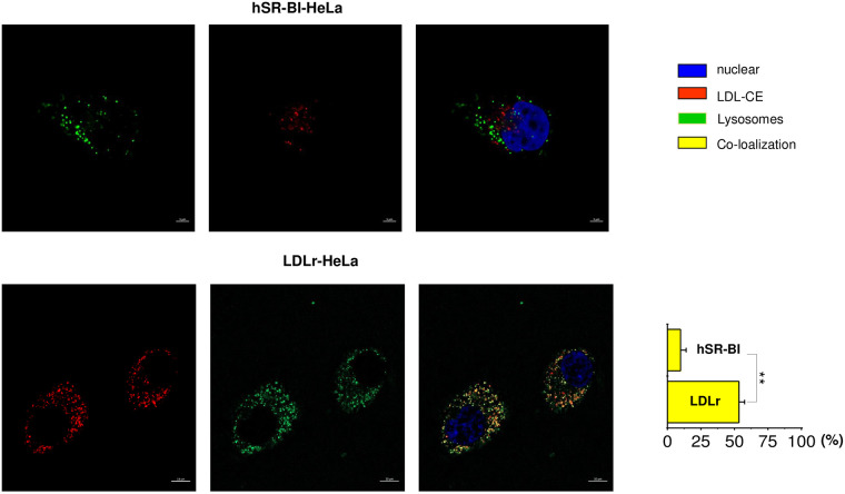
Fig 5. Transport of CE-LDL to the lysosomal compartment in various HeLa cells.
HeLa cells were plated at 30% confluency on glass covered slips and then pre-incubated in serum free media for 24 hours prior to experiments. Labelled CE-LDL were added to the media for 1 hour, washed and chased for 30–60 minutes in serum/lipoprotein free media. In the presence of Lysotracker Green (lysosomal marker). The data is arranged in rows with a color code seen on the right side of the panels. The confocal images of CE labeled LDL internalization to lysosomes utilizing Lysotracker green are shown for mock, SR-BI and LDLr HeLa cells as indicated at the top of the panels. The bar corresponds to 10 μm, * p < 0.015, ** p<0.01.

