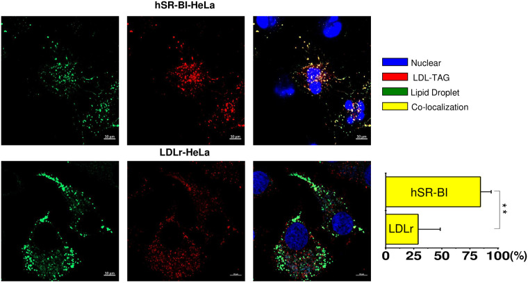
Fig 7. Co-localization of TAG labeled LDL with neutral BP -stained LDs.
HeLa cells were plated at 30% confluency on glass cover slips and pre-incubated in serum free media for 24 hours prior to experiments. Labelled LDL were added to the media for 1 hour, washed and chased for 60 minutes in serum/lipoprotein free media. Cells were stained for LDs utilizing Neutral BODIPY as described in Materials and methods. The confocal images of -TAG labeled LDL co-localized with neutral BP-stained LDs are demonstrated for various receptor expressing HeLa cells. The color code and extent of co-localization are shown in the right columns. The cell type is specified at the top of the figure. The bar corresponds to 10 μm, ** p < 0.01.

