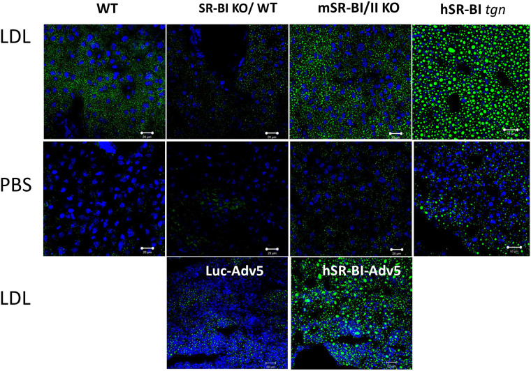
Fig 12. LDL-lipid accumulation in the livers of various mice.
Mice were fasted overnight and injected IV with either PBS (middle panel) or 2 mg of human LDL (upper and lower panels) and sacrificed 2 hours later. The upper two rows of panels show LD staining in liver cryosections from normal, SR-BI/II KO, hSR-BI transgenic SR-BI/II KO and hSR-BI transgenic normal mice after (+) or without (-) an LDL injection. The lower panels show LD staining in liver cryosections of Luc-AdV5 and SR-BI-AdV5 treated SR-BI KO mice following LDL injection. The animal codes are written on the panels. The bar corresponds to 20 μm.

