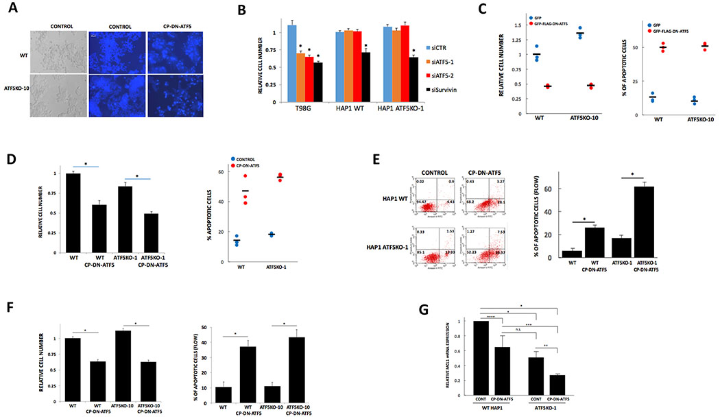
Figure 1.
HAP1 cells lacking active ATF5 respond to DN-ATF5, indicating that ATF5 is not the obligate target for DN-ATF5. A, Photomicrographs of WT and ATF5KO-10 HAP1 cells in bright field (left) or stained with Hoechst 33342 under control conditions (center) or after exposure to 100 μM CP-DN-ATF5 for 3 days in (right). B, ATF5 knockdown does not affect survival of HAP1 or HAP1 ATF5KO-1 cells, but does kill T98G cells. In contrast survivin knockdown compromises survival of all 3 cell types. Cultures were transfected with indicated siRNAs and 3 days later were evaluated for relative cell number. Values are means ± SEM. For T98G cells, data are from 5 independent experiments, total N=19-20 per condition. For HAP1 WT cells, data are from 5 independent experiments, total N=16 per condition. For HAP1 ATF5KO-1 cells, data are from two independent experiments, total N=10 per condition. *P<0.001 compared with siCTR for each cell type. C, FLAG-GFP-DN-ATF5 transfection reduces survival of WT and ATF5KO-1 HAP1 cells. Replicate cultures (N=3 per condition) were transfected as indicated and assessed 3 days later for relative numbers of transfected (GFP+) cells (left panel) and for proportion of transfected cells with apoptotic nuclei (right panel). D, CP-DN-ATF5 treatment reduces survival of WT and ATF5KO-1 HAP1 cells. Replicate cultures as indicated were treated without or with 100 μM CP-DN-ATF5 and assessed 3 days later for total cell number (left panel) and proportion of cells with apoptotic nuclei (right panel). For left panel, values represent means ± SEM for 3 independent experiments, each carried out in triplicate. *P<0.001. Right panel data are from one experiment carried out in replicate cultures. E, CP-DN-ATF5 treatment reduces survival of WT and ATF5KO-1 HAP1 cells as determined by flow cytometry. Left panel shows example of flow cytometric analysis of cell death in HAP1 WT and HAP1 ATF5KO-1 cultures treated with or without 100 μM CP-DN-ATF5 for 3 days. Right panel shows quantification of apoptotic cells with values representing means ± SEM for 2 independent experiments, each carried in triplicate (total N=6). *P<0.001. F, CP-DN-ATF5 treatment reduces survival of WT and ATF5KO-10 cells. Replicate cultures of WT and ATF5KO-10 cells as indicated were treated without or with 100 μM CP-DN-ATF5 and assessed 3 days later for cell number (left panel) or for % of apoptotic cells by flow cytometry. Values for cell numbers represent means ± SEM for 3 independent experiments, each carried out in triplicate. *P<0.001Values for % apoptosis represent means ± SEM for 4 independent experiments, each carried with 2-3 replicates (total N=10). *P<0.001. G, MCL1 mRNA levels are reduced in ATF5KO-1 cells compared to WT HAP1 cells and CP-DN-ATF5 lowers MCL1 mRNA levels in both cell types. Indicated cells were treated with or without 100 μM CP-DN-ATF5 for 3 days and assessed for MCL1 mRNA levels. Data represent mean values ±SEM and are from 9 independent determinations. *P<0.001; **P=0.01; ***P=0.02; ****P=0.03.

