
Fig. 1.
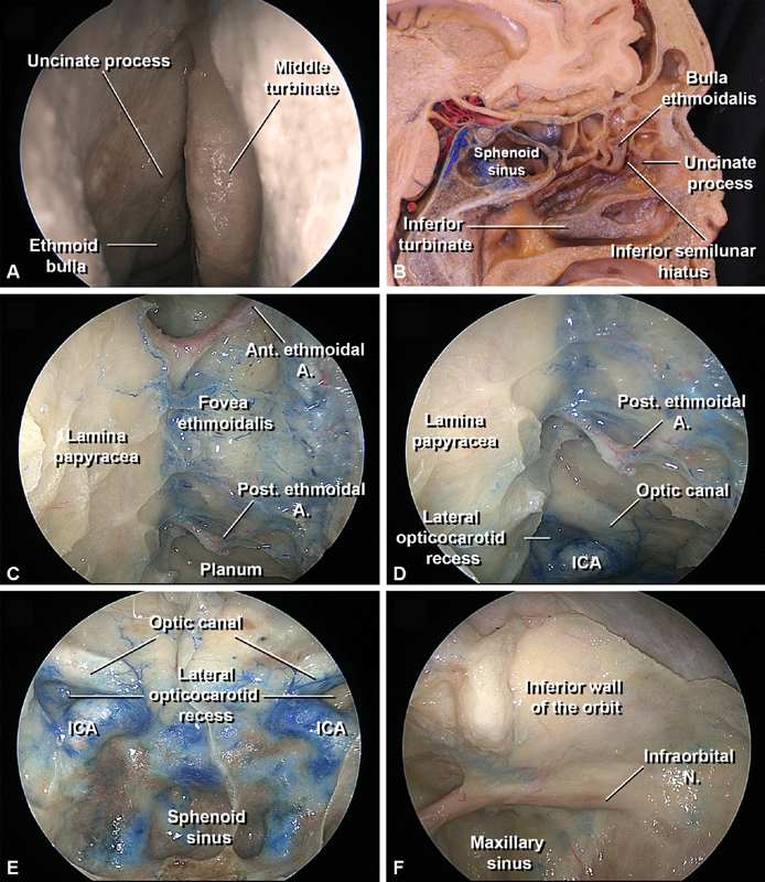
Medial and inferior anatomical relationships of the orbit. The orbit from an endoscopic endonasal perspective, 0-degree endoscopic view. ( A ) Identification of the right ethmoid bulla and uncinate process in the middle nasal meatus after careful medial displacement of the middle nasal turbinate. ( B ) Sagittal dissection of the left nasal cavity, the middle turbinate has been completely removed. The bulla ethmoidalis and the uncinate process have been partially divided. The space between the uncinate process and the bulla is the inferior semilunar hiatus. ( C ) The anterior and posterior ethmoidal canals with the respective arteries are visualized at the superior edge of the lamina papyracea and cranial base after removal of the ethmoidal cells. These canals divide the anterior fossa floor and nasal cavity roof into frontal (anterior to the anterior ethmoidal canal), cribriform (between both canals), and planum areas (posterior to the posterior ethmoidal canals), which approximately correspond to the to the bulbar, retrobulbar, and apical parts of the orbit. ( D ) Relationship of the posterior ethmoidal canal with the orbital apex posteriorly: the optic canal prominence is located superior to the lateral opticocarotid recess, a triangle-shaped depression superolateral to the carotid prominence. The lateral opticocarotid recess corresponds to the optic strut base intracranially, and the clinoid segment of the internal carotid artery is located medially. ( E ) Endonasal view of the posterior wall of the sphenoid sinus. ( F ) The inferior wall of the orbit corresponds to the superior wall of the maxillary sinus. The infraorbital nerve crosses through the infraorbital canal until it exits via the infraorbital foramen located in the inferior orbital rim. ( G ) Close view of the lamina papyracea. The medial wall of the maxillary sinus has been removed but the underlying mucosa has been preserved. ( H ) The lamina papyracea has been resected exposing the periorbita and the mucosa of the medial wall of the maxillary sinus has been removed. ( I ) The periorbita underneath the lamina papyracea has been incised and the orbit fat has been removed, exposing the neurovascular orbital contents through the medial orbit wall. ( J ) The superior wall of the maxillary sinus has been removed medially to the infraorbital nerve and the periorbita has been incised, exposing the neurovascular orbital contents through the medial and inferior orbit walls. Note how the infraorbital branch of the internal maxillary artery follows the infraorbital nerve at the infraorbital groove and receives an anastomosis from the ophthalmic artery. ( K ): Detailed view of the orbital apex and the optic canal. ( L ) Specimen divided in the sagittal plane, the ethmoid labyrinth and the medial wall of the maxillary sinus have been removed exposing the infraorbital canal and the orbital plate of the ethmoidal bone (lamina papyracea). A., Anterior; ICA, internal carotid artery; M, muscle; N, nerve; Post, posterior.