
Fig. 3.
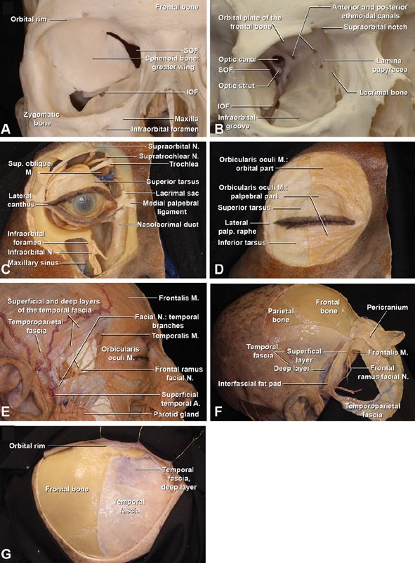
Anterior anatomical relationships of the orbit. ( A, B ) A anterior and oblique overview of the osseous components of the right orbit, which is formed by seven different bones: the frontal, zygomatic, sphenoid, lacrimal, ethmoid, palatine bones, and maxilla. Note the paper-thin consistency of the lamina papyracea, and its relationship with the anterior and posterior ethmoidal canals, within the superior edge of the lamina papyracea and the medial edge of the orbital plate of the frontal bone. The canals offer exit to the anterior and posterior ethmoidal arteries (which are branches of the ophthalmic artery) and the nasociliary nerve (division of the ophthalmic nerve). The nasolacrimal groove is formed anteriorly by the frontal process of the maxilla and posteriorly by the lacrimal bone. The optic canal is observed close to the posterior ethmoidal canal and contains the optic nerve, ophthalmic artery and sympathetic nerve fibers. The SOF and IOF are identified. Note how the lower end of the SOF blends inferiorly with the posterior end of the IOF. Note the infraorbital foramen of the anterior maxilla and the supraorbital notch at the supraorbital rim. ( C ) Oblique view of the right orbit. The skin surrounding the orbit, extraocular muscles and orbital fat have been removed, and the maxillary sinus opened. The medial and lateral canthus are demonstrated. The supra-orbital nerve emerges from the supra-orbital foramen and the supratrochlear nerve is located medially between the supraorbital foramen and the trochlea of the superior oblique muscle. The inferior orbital wall has been removed medially up to the infraorbital canal. The infraorbital nerve leaves the orbit through the infraorbital foramen and divides into the palpebral, nasal and superior labial branches, supplying sensory innervation of the skin of the lower eyelid, medial cheek, lateral nose and upper lip, and the mucosa of the anteroinferior nasal septum and oral mucosa of upper lip. ( D ) Right anterior view of the orbit after skin removal. The orbital and palpebral portions of the orbicularis oculi muscle are observed in the medial half of the orbit. The lacrimal part of the orbicularis oculi muscle lies underneath these structures, extending behind the lacrimal sac and attaching to the lacrimal bone. The orbicularis oculi muscle surrounds the orbital rim circumferentially and extends into the lids, temple and cheek, having its fibers interdigitated with the occipitofrontalis and the corrugator muscles. On the lateral side of the orbit, the orbicularis oculi muscle has been resected and the two dense plates of connective tissue know as tarsi are observed. ( E ) Right lateral view to illustrate the course and location of the temporal branches of the facial nerve. The temporal division of the facial nerve emerges directly through the parotid gland and divides in three rami: anterior, middle and posterior. The temporal branches are located superficial to the temporoparietal fascia (also known as superficial temporal fascia). The middle ramus (frontal ramus), which innervates the frontalis muscle, is located anteroinferior to the frontal branch of the superficial temporal artery. The frontalis muscle is continuous laterally with the temporoparietal fascia and is medial to the superior temporal line. ( F ) Dissection of the right temporal area. The temporoparietal fascia and the frontalis muscle have been reflected anteriorly and part of the temporoparietal fascia has been resected to expose the frontal ramus of the facial nerve and the superficial temporal artery that run on this layer. The temporal fascia is continuous with the pericranium medially and divides 2 to 3 cm above the zygomatic arch into two layers, superficial and deep. In between these layers the interfascial fat pad is encountered. To protect the frontal ramus of the facial nerve the dissection of the skin flap is performed between the deep and superficial layers of the deep temporal fascia (interfascial dissection) or under the deep layer (subfascial dissection). ( G ): Right frontotemporal exposure within trifacial temporal dissection. The interfascial–subpericranial flap has been folded anteriorly. A, artery; IOF, inferior orbital fissure; M, muscle; N, nerve; Palp, palpebral; SOF, superior orbital fissure.