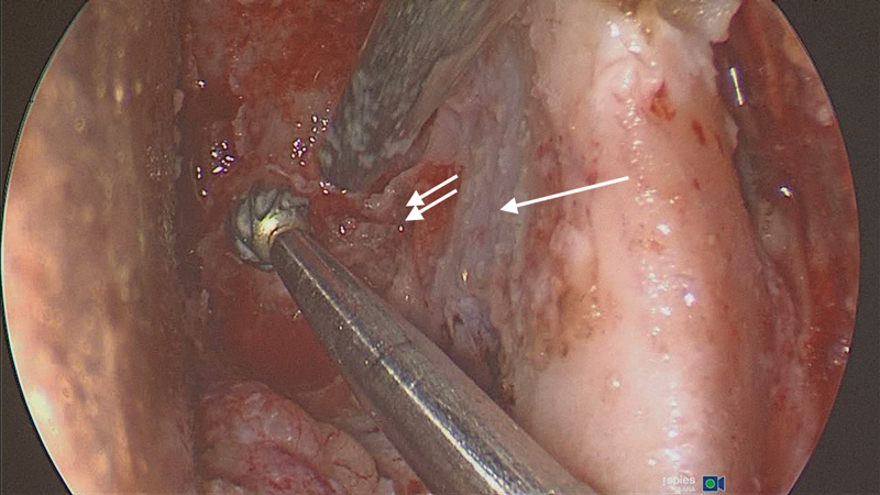
Fig. 10.

The lateral orbital corridor: thick area of hyperostotic bone requiring removal to expose the temporalis muscle anterolaterally (arrow), the middle cranial fossa posteriolaterally (double arrow) and the anterior cranial fossa dura superiorly (Left eye).
