Fig. 8.

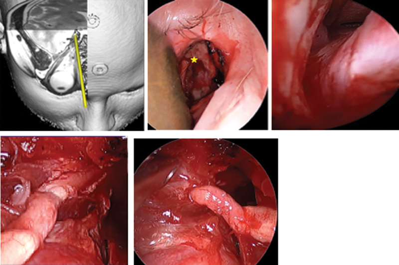
Optic nerve visualized through right medial (precaruncular) approach. Intraoperative appearance, right orbit, and computer planning image demonstrating trajectory. A 67-year-old male with history of choroidal melanoma 9 years prior treated with brachytherapy developed recurrence and underwent enucleation. Pathology showed tumor extending to optic disk, posterior globe, and subarachnoid space. Nodular areas of enhancement along the optic nerve were noted on imaging, so he was referred for endoscopic resection of the optic nerve and postoperative proton beam radiotherapy. The nerve was transected at the distal end of its intracranial course. Pathology demonstrated no further melanoma, and the patient was able to retain the same functional enucleation prosthesis. ( A ) Planning illustration of precaruncular approach. ( B ) Right precaruncular approach, superficial view. A malleable retractor is displacing orbital contents laterally. The anterior ethmoid artery ( star ) marks the frontoethmoid suture, which corresponds to the skull base. ( C ) Right optic nerve at the orbital apex exiting the optic canal. ( D ), Medial view of the optic nerve with the annulus removed and the optic canal decompressed. ( E ) Lateral aspect of the mobilized optic nerve entering the annulus prior to removal.
