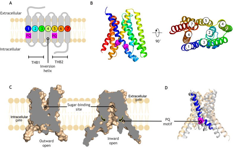
Fig. 2.
Crystal structure of the SWEET sugar transporter. (A) The eukaryotic SWEET transporter contains seven transmembrane alpha helices, which can be split into two three-helical bundles (THBs) (PDBe entry: 5ctg). The PQ sequence motif is located in the first helix of each THB, suggesting the full-length protein evolved by gene duplication. The fourth helix links the two THBs together and is often referred to as the inversion helix. (B) Crystal structure of the eukaryotic SWEET transporter coloured from N-terminus (blue) to C-terminus (red). The two PQ-motifs are highlighted and shown in space-filling representation (magenta). Right panel – view rotated 90°; helices are labelled. (C) Alternating access transport mechanism as revealed from crystal structures of the bacterial semiSWEET transporters (PDBe entries: 4×5n, 4×5m). The central sugar-binding site is labelled. Arrows indicate structural changes upon sugar binding. (D) The crystal structures of the outward- and inward-facing semiSWEET transporters (grey and beige, respectively) have been superimposed. Helix 1 has been coloured (blue) with the PQ-motif highlighted (magenta).

