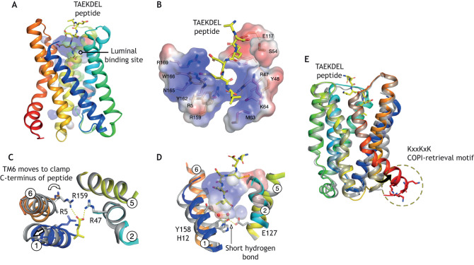
Fig. 4.
Mechanism of receptor activation. (A) Crystal structure of the KDELR bound to the TAEKDEL peptide (PDBe entry: 6i6h). The electrostatic surface of the peptide-binding site is shown. (B). Close up view of the peptide-binding site shown in A. The TAEKDEL peptide is shown in sticks (yellow), with key binding site side chains highlighted (wheat). (C) Top-down view of the peptide-binding site, showing the structural changes accompanying peptide binding. (D) Close-up view of the peptide-binding site shown in A, displaying the H2O molecules bound at the base of the pocket. (E) Comparison of the inactive (grey) and activated (coloured) receptor. The key structural change of the cytoplasmic side of the receptor is highlighted.

