Fig. 1.
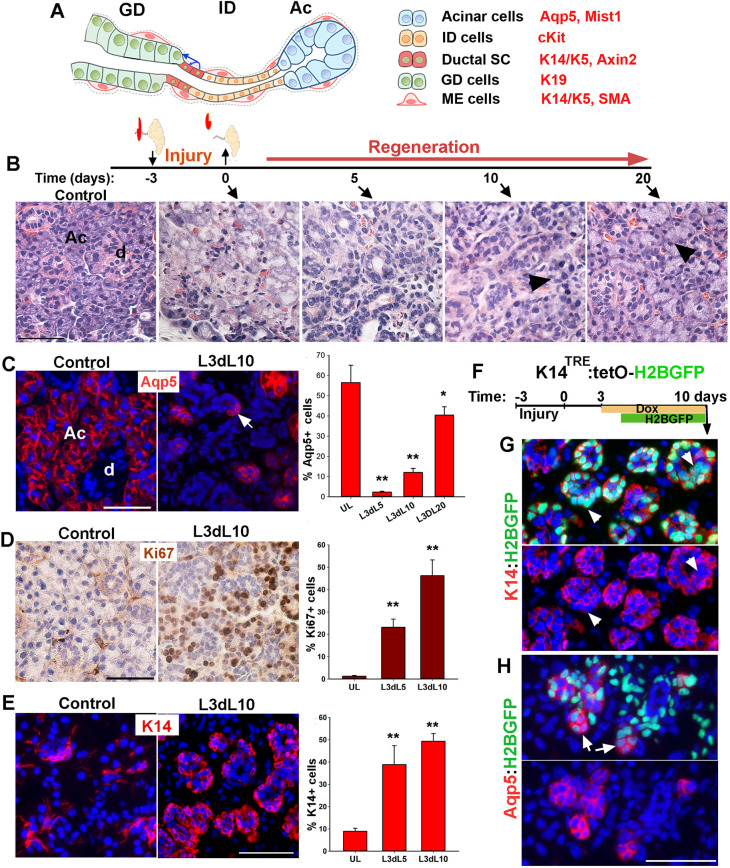
Characterization of salivary gland regeneration in a model of severe injury. (A) Schematic representation of secretory complex in mouse SMG and specific markers used to identify each cell type. Ac, acini; ID, intercalated duct; GD, granular duct. (B) Timeline of duct ligation/deligation injury and images of Hematoxylin and Eosin-stained sections of SMG at the indicated time points. Arrowheads indicate acini. (C) Distribution and quantification of Aqp5+ cells in control (UL) and injured gland collected at 5, 10 and 20 days post-de-ligation (L3dL). Arrow indicates proacinar cells budding from tubular structures. d, duct. (D,E) Distribution and quantification of Ki67 proliferation marker and K14 at days 5 and 10 post-ligation when tubular structures are present. Image shown is from day 10 post-de-ligation (L3dL10). Data are mean±s.e.m (n=3 females), one-way ANOVA and post-hoc Tukey's test (*P<0.05 and **P<0.001). (F) Timeline of nuclear tracking of K14-H2BGFP. (G,H) Images of regenerative gland stained for K14 or Aqp5. Arrowheads in G indicate nuclear GFP in K14neg cells. Arrows in H indicate acini-like structures with dim green fluorescence in their nuclei. Images are representative of n=3 female mice. Scale bars: 50 µm.

