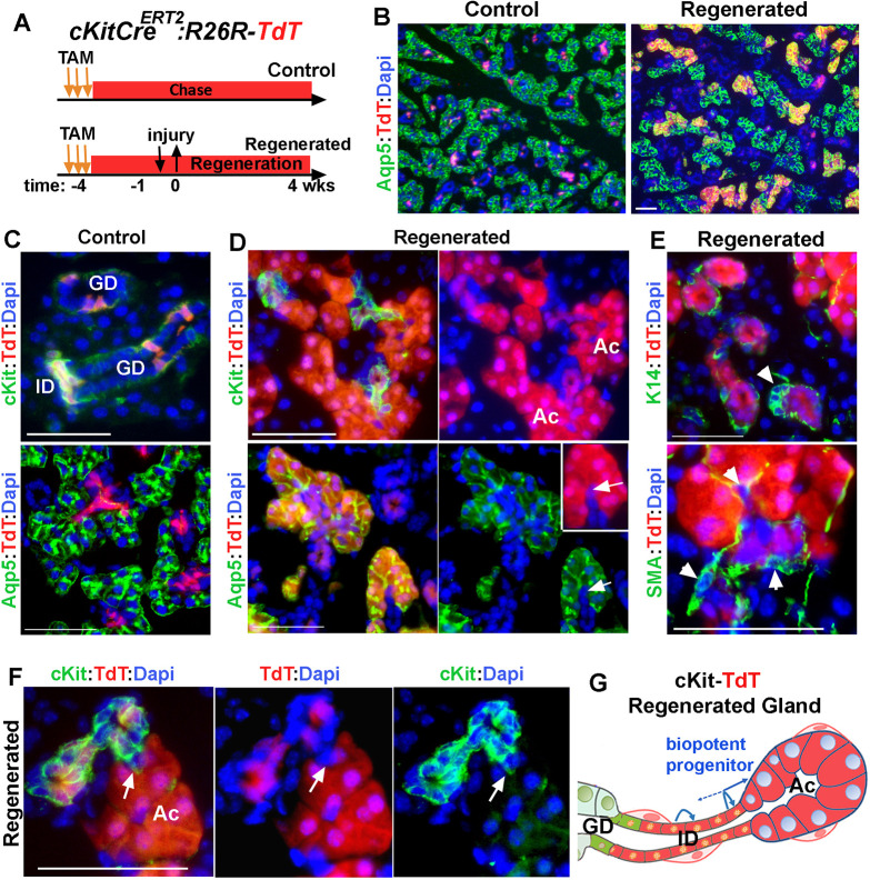
Fig. 6.
Contribution of Kit+ ductal cells to regeneration of acini. (A) Timeline of label induction and lineage tracing of Kit+ cells during injury and regeneration. (B-D) TdT-lineage traced cells in control and regenerated glands stained for Aqp5 or Kit. Arrows in D indicate a single TdT+ ID cell contiguous with TdT+ acini. (E) Images of TdT-labeled cell clusters stained for K14 and SMA. Arrowheads indicate ductal stem cells. (F) A small number of TdT-labeled acini are not contiguous with TdT-labeled Kit+ cells (arrow). Nuclear blue staining is DAPI. ID, intercalated duct; AC, acini; GD, granular duct. Scale bars: 50 µm. Images are representative of three glands from female mice. (G) Summary of results of lineage tracing of ID cells, indicating dedifferentiation of Kit+ cells to a bi-potent progenitor cell population that re-differentiated to both ID and acinar cells. Distal ID cells may self-replicate.

