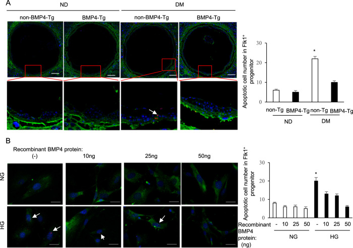
Fig. 4. BMP4 overexpression block Flk-1+ progenitor apoptosis in vivo and in vitro.
a Representative TUNEL staining images showing apoptotic cells (red dots) in Flk-1+ progenitors (green) in yolk sac. Cell nuclei were stained with DAPI (blue), Scale bar = 100 μm. Quantification of TUNEL positive in Flk-1+ progenitors was shown in the graph. B: Representative TUNEL staining images showing apoptotic cells (red dots) in Flk-1+ progenitors (green) treated with different concentrations of recombinant BMP4 protein. Cell nuclei were stained with DAPI (blue), Scale bar = 50 μm. Quantification of TUNEL positive in Flk-1+ progenitors was shown in the graph. Experiments were performed using three embryos from three different dams per group (n = 3); * indicates significant difference compared to the other groups (P < 0.05) in one-way ANOVA followed by Tukey tests. ND: nondiabetic; DM: diabetes mellitus. NG: normal glucose; HG: high glucose.

