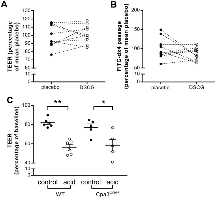
Figure 7.
Acid-induced impairment of the duodenal mucosal barrier is not mediated by mast cells. Mucosal barrier function after acid perfusion following treatment with placebo (black dots) and DSCG (white dots) was evaluated in Ussing chambers by measuring TEER (A) and passage of FITC-dx4 (B). n = 10 for both groups. (C) Acid-induced reduction in TEER in wild type and in deficient mast cell Cpa3Cre/+ mice. Results are expressed relative to the mean of the control group. DSCG, disodiumcromoglycate; FITC-dx4, fluorescently labeled dextran of 4 kDa; TEER, transepithelial electrical resistance; WT, wild type.

